Elbow Fractures: Supracondylar Humerus

What is the elbow?

The elbow is a joint in the arm that connects three bones: (Figure 1)

  • Humerus: bone in upper arm stretching from the shoulder to the elbow
  • Radius: forearm bone stretching from the elbow to the wrist on the thumb side
  • Ulna: forearm bone stretching from the elbow to the wrist on the pinky side

These three bones allow the arm to bend, straighten, and rotate. The radius allows the forearm to rotate the palms up or down. The ulna acts as a hinge, sitting in the pocket of the end of the humerus, to allow bending and straightening of the arm.

What causes supracondylar humerus  fractures?

Because the elbow is not well protected by soft tissue and experiences high impact trauma during falls, elbow fractures are very common in children, accounting for about 10% of pediatric fractures. Supracondylar humerus fractures are the most common elbow fracture seen in children.  Elbow fractures most commonly occur in children between the ages of 5-7 during a fall on an outstretched hand.

Signs and Symptoms

Symptoms of supracondylar humerus fractures may include:

  • Pain in the elbow
  • Difficulty bending or straightening the arm
  • Swelling or bruising in the elbow
  • Visible deformity (Figure 2)

Diagnosis of Supracondylar Humerus Fractures:

If an supracondylar fracture is suspected based on the symptoms above, the child should be brought to the emergency room immediately for physical examination and imaging tests (X-ray).

During physical exam, an orthopedic doctor will carefully inspect the arm, and test for nerve and blood vessel involvement.

Typically, X-ray imaging is sufficient to confirm the diagnosis of a supracondylar humerus fracture.

Rarely, the doctor may order a CT or MRI in order to visualize the fracture and associated nerves and blood vessels.

Treatment of Elbow Fractures:

Treatment plans for elbow fractures vary based on the type and location of the fracture. A Campbell Clinic orthopedic physician will address the findings from physical exam and imaging studies (X-ray) to determine the best treatment plan for the fractured elbow.

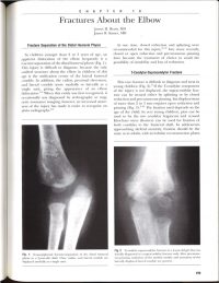
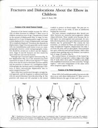
Many mild fractures can be treated with a short period of casting (3-4 weeks) especially in yonger patients.  Fractures in which the bone fragments have displaced usually require surgical intervention and placing metal pins to reattach the bone fragments.  (Figure 3)  Many times these pins can be inserted without making an actual incision. (Figure 4).  Those rare fractures in which the blood supply to the arm is affected are true emergencies and need to be evaluated immediately. (Figure 5).

Outlook (Prognosis):

Depending on type, severity and treatment plan, supracondylar humerus fractures can take up to 6 to 8 weeks to completely heal. Some fractures may heal faster (non-surgical).  Most children who sustain supracondylar humerus fractures do very well long term and completely heal.

Elbow stiffness and forearm weakness are common after removing a cast, but proper physical therapy and strengthening exercises are very effective in regaining full strength and mobility.

It is recommended to schedule follow up appointments with an orthopedic physician every few weeks until the elbow has fully healed.  In more severe fractures, X-ray imaging is recommended every few weeks until the bones have fully healed.

In order to avoid serious complications, it is very important to seek medical treatment as soon as an elbow fracture is suspected.

Published Articles

For appointments call
901-759-3111